
Longitudinal hippocampal atrophy was tied to cognitive decline independently of amyloid-beta or tau in clinically normal older adults, suggesting that non-Alzheimer’s pathologies may contribute to hippocampal-mediated cognitive decline.
Faster hippocampal atrophy was correlated with faster cognitive decline (R2=0.28, P<0.0001), reported Bernard Hanseeuw, MD, PhD, of Massachusetts General Hospital in Boston, and co-authors.
On its own, hippocampal volume slope accounted for 10% of the variance in cognitive decline, the researchers reported in Neurology. About half the variance in cognitive decline was explained by combining longitudinal changes in imaging biomarkers of amyloid, tau, brain volume, and cerebrovascular disease.
“As the first disease-modifying therapies for Alzheimer’s disease are coming to clinical practice, understanding the causes of cognitive decline as early as possible is critically important,” Hanseeuw said.
“To this end, amyloid imaging allows disclosing Alzheimer’s disease pathology in individuals who are not yet experiencing cognitive impairment. However, amyloid imaging does not allow disclosing other neuropathologies that are frequent in the elderly population,” he told MedPage Today.
“By monitoring not only amyloid, but also neocortical tau deposition using PET and hippocampal atrophy using MRI, we demonstrated that these biomarkers explained more than half the variance in cognitive decline in a sample of 128 clinically normal individuals followed during a decade,” Hanseeuw continued.
“Progressive hippocampal atrophy in the absence of tau accumulation explained 10% of the cognitive variance, suggesting LATE [limbic-predominant age-related TDP-43 encephalopathy] was present in some individuals from our sample, a condition that is unlikely to improve with anti-amyloid therapies,” he pointed out.
“In practice, measuring tau and hippocampal volume in addition to amyloid biomarkers is useful to better ascertain the etiological diagnosis of patients presenting with cognitive decline,” he added.
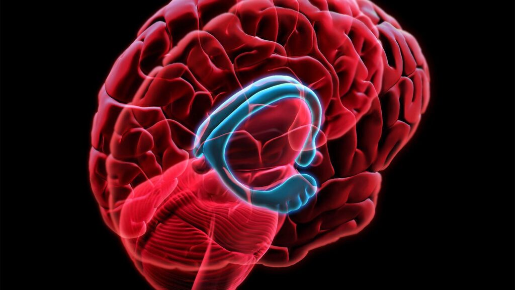
Hippocampal volume atrophy is a well-known biomarker of memory impairment, but is less specific than amyloid or tau imaging for Alzheimer’s pathology. “This lack of specificity could provide indirect information about potential co-pathologies that cannot be observed in vivo,” the researchers noted.
Hanseeuw and co-authors followed 128 older adults without dementia or overt cognitive impairment in the Harvard Aging Brain Study to assess relationships between longitudinal imaging and cognitive decline. Median age at study inclusion was 73, and 56% were women.
The researchers prospectively collected serial MRI, neocortical amyloid imaging with Pittsburgh compound B (PiB) PET, and entorhinal and inferior temporal tau with flortaucipir PET. About a quarter (27%) of participants showed an initial high-amyloid burden on PET imaging.
Cognition was monitored throughout the study using the Preclinical Alzheimer Cognitive Composite (PACC), an average of z-scored performances on five tests sensitive to cognitive decline. Over a median follow-up of 7.3 years, participants had a median of eight annual cognitive evaluations.
In total, 48% of the variance in PACC decline was explained by changes in biomarkers. “Only 2% of the variance in cognitive changes was still explained by age at baseline after taking into account biomarker changes, indicating that most of the age-related cognitive decline was explained by this model,” the researchers noted.
The results were partly driven by a few participants who progressed to mild cognitive impairment or dementia during the study. “Removing these individuals from the analyses did not modify the results other than reducing the total amount of cognitive variance explained to 31%,” Hanseeuw and co-authors observed.
The study was not designed to detect atypical presentations of Alzheimer’s disease or non-Alzheimer’s pathologies, they acknowledged. “Group studies may be insensitive to atypical patterns of biomarker progression, and larger studies are required to evaluate inter-individual variations in biomarkers trajectories,” they added.
-
Judy George covers neurology and neuroscience news for MedPage Today, writing about brain aging, Alzheimer’s, dementia, MS, rare diseases, epilepsy, autism, headache, stroke, Parkinson’s, ALS, concussion, CTE, sleep, pain, and more. Follow
Disclosures
This research was supported by the NIH, the Belgian Fund for Scientific Research, and the Queen Elizabeth Medical Foundation.
Hanseeuw reported funding from the Belgian Fund for Scientific Research. Co-authors reported relationships with the NIH, Alzheimer’s Association, International Society to Advance Alzheimer’s Research and Treatment, the Journal of Alzheimer’s Disease, Biogen, Humana Healthcare, Leerink Partners, Expert Connect, Eisai, Eli Lilly, Genentech, Analysis Group, Celgene, Verily Life Sciences, Merck-Serono, Novartis, and Genzyme.
Primary Source
Neurology
Source Reference: Hanseeuw BJ, et al “Association of pathological and volumetric biomarker changes with cognitive decline in clinically normal adults: Harvard Aging Brain Study” Neurology 2023; DOI: 10.1212/WNL.0000000000207962.
Please enable JavaScript to view the